Denoising Label-free Two-Photon Microscopy Images using Deep Learning
What is Cervical Precancer?
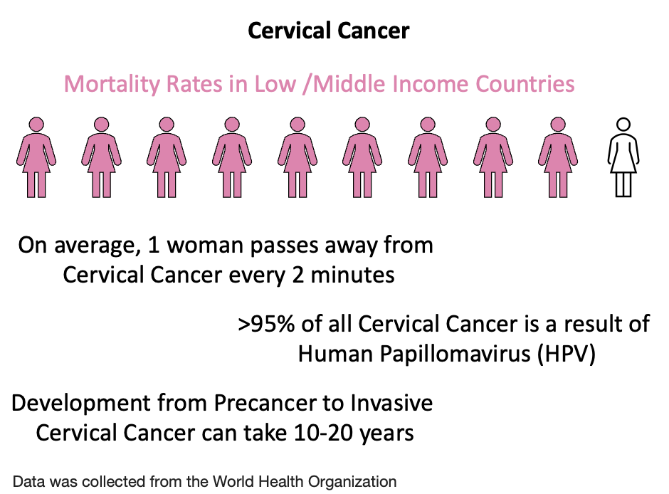
Globally, Breast and Cervical cancer are two of the leading forms of cancer in women.
For Cervical cancer, high HPV vaccination rates and access to efficient treatments has
led to low incidence and mortality rates in high income countries, however, 9/10
cervical cancer death originate from low-income countries.
Cervical cancer is the fourth most common cancer in women worldwide.
Cervical cancer is characterized by neoplastic growth of cervical cells
at the transformation zone of the endocervix and ectocervix1. Cervical
precancer originates from persistent infection with human papillomavirus
(HPV)1-3. HPV can induce cellular transformations which can lead to
dysplasia and cervical precancer but does not always result in precancer
development1,2. Cervical precancer is traditionally screened for using a
cytology-based Papanicolaou (Pap) smear exam and molecular-based HPV DNA
test1-3. In a Pap smear test, cells are collected from the cervix using
a spatula or brush and imaged using microscopy for abnormal cells3. If
abnormal cells and oncogenic HPV DNA are detected, patients undergo further
colposcopic examination3. Colposcopy involves low magnification imaging of
the cervix for dysplasia and collection of biopsies from the abnormal regions
of the cervix3. Biopsies are used to grade the lesion and determine
pre-cancerous status. If the biopsy is identified as pre-cancerous,
ablative (burning/freezing) or excisional (surgical excision) treatment
methods are used to remove the precancerous lesions3.
Limitations in Clinical Nonlinear Optical Imaging
Most clinical TPEF imaging has been conducted on skin with some endoscopic TPEF imaging13-17.
A key consideration in clinical TPEF imaging is fast image acquisition of high SNR data. In
preclinical studies, excised biopsies are plated on glass bottom dishes and imaged using a
high NA objective in a lab without major consideration of maximum permissible light dose and
motion artifacts18. Comparatively, in vivo imaging is expected to have motion artifacts from patients
and reduced irradiation parameters for safety, requiring high-speed imaging and lower illumination
powers. These two factors can significantly impact the acquired image SNR.
Current in vivo MPM imaging systems include the DermaInspect, MPTflex, and MPTcompact systems.
These systems utilize high NA=1.3 objectives to capture label-free TPEF images in patients19.
However, to accommodate the high NA objective, the systems are designed to be large and bulky,
limiting their use for clinical acquisition to external skin imaging. A micro-endoscope MPM system
with a NA=0.8 objective is also available but is limited by its collection optics that prevent
internal body use20. As such, hardware improvements are still needed for endoscopic MPM imaging.
Further, software improvements are required to recover image quality from low SNR images to
account for the reduced illumination powers and potential motion artifacts.
In this project, we will focus specifically on software improvements for biologically relevant
image restoration of predictive metabolic metrics as an initial step. We aim to demonstrate the
recovery of image quality and morphofunctional metrics of metabolic activity using DL, which can
aid in the clinical translation of MPM.
What is Two-Photon Excited Fluorescence Microscopy?
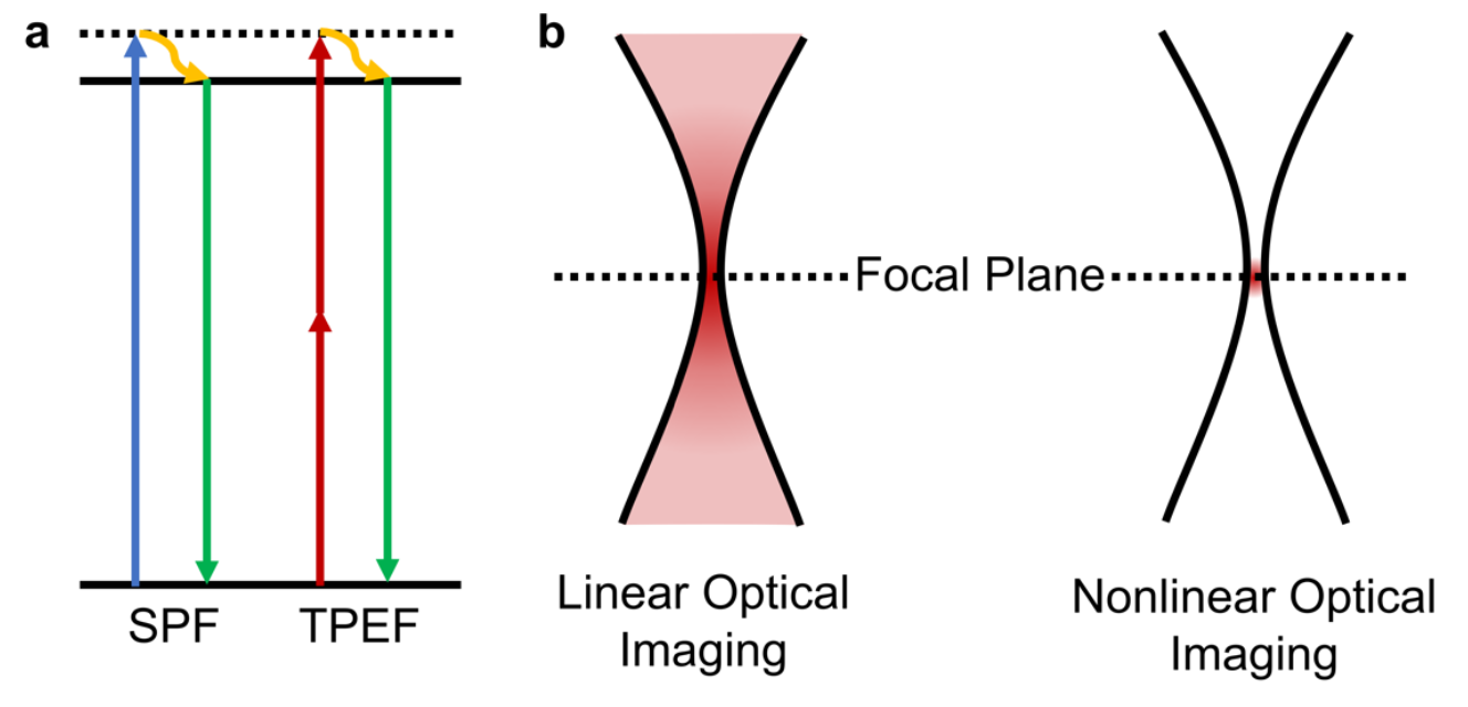
Figure 1: (a) Jablonski diagram for single photon and two-photon excited fluorescence (b) Visual representation of fluorescence intensity along the axial plane for SPF and TPEF.
Two-Photon excited fluorescence microscopy in a nonlinear optical imaging technique.
Nonlinear optical (NLO) imaging exploits nonlinear light matter interactions that
generally occur only at the focal plane, providing intrinsic depth sectioning4-6.
Multiphoton microscopy (MPM) is an NLO imaging technique that leverages the simultaneous
absorption of two or more low energy photons that arrive “simultaneously” at the fluorophore7 .
Owing to the “simultaneous” absorption of low energy photons, the fluorophore achieves a greater
energy state5,7. After a short period of time, the molecule returns to its ground state;
however, due to the higher energy state from multiphoton excitation, the emitted fluorescence is of a
lower wavelength compared to the excitation wavelength5,7.
To generate nonlinear optical events, high-powered, ultrafast pulsed lasers
(on the order of femtoseconds) are used8. Multiple nonlinear optical processes
can be generated using these lasers; however, here, we focus specifically on Two-Photon
excited fluorescence (TPEF).
TPEF is a process in which two photons are absorbed by atoms within a time window of
an attosecond (10-18 s), leading to the molecule reaching an excited electronic state9,10.
Quantum mechanically, a single photon is absorbed, bringing the atoms to a virtual
intermediate state before eventually reaching the final excited state by absorbing
a second photon9. Comparatively, in single-photon fluorescence (SPF), a single photon is
absorbed, causing atoms to reach their final excited state (Figure 1)9. After reaching
the final excited state, vibrational relaxation is observed in both SPF and TPEF before
the atoms return to their ground state, emitting a photon (fluorescence).
Another feature of TPEF is the highly confined three-dimensional excitation volume.
In SPF, excitation occurs along the entire axial plane constantly9. However, in
TPEF, nonlinear excitation needs high photon flux (1020-1030 photons/cm2·s) which
is only satisfied near the focus (Figure 1)9,10. This feature of TPEF enables
intrinsic depth sectioning and minimized photobleaching in out-of-focus regions.
The final advantage of TPEF compared to SPF is that TPEF typically uses NIR wavelength
laser light sources compared to visible wavelength lasers used in SPF. The absorption and
scattering of light by tissue are greater in the visible wavelength regime, minimizing the
potential penetration depth of light into bulk tissue. NIR light enables deeper penetration
of light by reducing the number of scattering events and absorption of the low energy photons9.
Simultaneously, TPEF mimics behaviors of higher energy SPF allowing for the excitation of
endogenous fluorophores that typically require high-energy photon excitation. Further, since a
NIR illumination source is used to excite a fluorophore with emission in the visible region,
greater separation in excitation and emission bands exist, leading to a higher SNR signal9.
Sources of Endogenous Contrast for Nonlinear Optical Imaging
A variety of endogenous biomolecules can be visualized using TPEF in biological samples. These
fluorophores include Keratin, Retinol, LipDH, NAD(P)H, FAD, Melanin, Lipofuscin, and electron-transferring
flavoprotein (ETF)4. Free, unbound FAD is a small portion of all FAD autofluorescence; the
majority of FAD signal is found within other flavoproteins like LipDH4. In the work described
in this project, FAD is used interchangeably to describe autofluorescence detected from
all flavin-associated proteins.
The most common sources of intracellular signal in TPEF are NAD(P)H and FAD4,6. Extracellularly,
collagen and elastin fibers contribute significant signals in TPEF due to crosslinking structures
within these proteins11,12. For all analyses of TPEF images in this project, NAD(P)H and
FAD signals are used, with collagen and elastin signals being masked. NAD(P)H is commonly excited
in TPEF using a 755 nm laser, and emission is collected at 460 ± 25 nm4,6. FAD is excited in TPEF
using a 860 nm laser, and emission is collected at 525 ± 25 nm4,6. These intrinsic contrast sources
have enabled the rapid translation of TPEF from the benchtop to clinical settings13-16.
To learn more about how these sources of endogenous contrast can be used for cancer diagnostics, look
at the link below.
Application of Deep Learning to NLO Images
Many ML and DL networks have been proposed for image restoration of biomedical images.
However, most proposed networks have been used to restore image quality in fluorescently stained samples. Exogenously
stained sample images feature higher SNR than label-free sample images owing to enhanced contrast from the fluorescence
stain. The same networks are not expected to have comparable performance on label-free images which have lower SNR.
DL-enabled label-free image restoration was first explored by Fast et al. (2020) to enhance image contrast and
extend the scanning area of a custom in vivo MPM exoscope imaging system21. Fast et al. (2020) demonstrated
using the content aware image restoration and enhancement (CARE) network sub-micron lateral resolution with a
large field of view in a fraction of the time compared to current commercial MPM imaging systems (DermaInspect)21.
However, no metrics on image quality or model performance were provided. More recently, Shen et al. (2022) presented
alternative methods for DL-enabled label-free image restoration of TPEF images22. Shen et al. (2022) demonstrated using
a modified generative adversarial network (GAN) ~4.5 dB improvement in peak SNR (PSNR) and 79% improvement in structural
similarity index measure (SSIM), two metrics used to assess the quality of a noisy image compared to a clean/ground
truth image22. However, tissue images were collected from thin optical sections (OS), featuring consistent SNR between
each image compared to bulk tissue, where SNR is expected to decay as a function of depth.
Shen et al. (2022) and Fast et al. (2020)
are the only studies to examine DL-enabled restoration of label-free TPEF
images21,22. Despite the observed improvement in image quality after restoration, there is a lack of understanding
regarding the impact denoising has on biomedical metrics extracted from these images. Morphofunctional metrics of
metabolic activity show immense potential for sensitive (93.3%) and specific (83.3%) detection of cervical precancer18.
It is therefore imperative for DL algorithms to demonstrate recovery of not only image quality but also biomedical metrics.
In this project, we cover some of our early efforts to understand the impact of DL-based image restoration on
metabolic metric recovery compared to standard image quality metrics and explore how these models could be used for
clinical diagnosis of cervical pre-cancer. To read our published work click on the link below.
References
Gates, A. et al. Screening for the prevention and early detection of cervical cancer: protocol for systematic reviews to inform Canadian recommendations. Syst. Rev. 10, (2021). https://doi.org/10.1186/s13643-020-01538-9/
World Health Organization. Screening and treatment of cervical pre-cancer. in Cervical Cancer Control: A Guide to Essential Practice 129–161 (2014).https://www.ncbi.nlm.nih.gov/books/NBK269601/
Thomas, G., Van Voskuilen, J., Gerritsen, H. C. & Sterenborg, H. J. C. M. Advances and challenges in label-free nonlinear optical imaging using two-photon excitation fluorescence and second harmonic generation for cancer research. J. Photochem. Photobiol. B Biol. 141, 128–138 (2014).https://doi.org/10.1016/j.jphotobiol.2014.08.025
Cho, H. J., Chun, H. J., Kim, E. S. & Cho, B. R. Multiphoton microscopy: An introduction to gastroenterologists. World J. Gastroenterol. 17, 4456–4460 (2011).https://doi.org/10.3748%2Fwjg.v17.i40.4456
So, P. T. C., Dong, C. Y., Masters, B. R. & Berland, K. M. Two-Photon Excitation Fluorescence Microscopy. Annu. Rev. Biomed. Eng. 2, 399–429 (2000).https://doi.org/10.1146/annurev.bioeng.2.1.399
Oheim, M., Michael, D. J., Geisbauer, M., Madsen, D. & Chow, R. H. Principles of two-photon excitation fluorescence microscopy and other nonlinear imaging approaches. Adv. Drug Deliv. Rev. 58, 788–808 (2006).https://doi.org/10.1016/j.addr.2006.07.005
Quinn, K. P. et al. Optical metrics of the extracellular matrix predict compositional and mechanical changes after myocardial infarction. Sci. Rep. 6, 1–12 (2016).https://doi.org/10.1038/srep35823
Chai, D. et al. Quantitative assessment of UVA-riboflavin corneal cross-linking using nonlinear optical microscopy. Investig. Ophthalmol. Vis. Sci. 52, 4231–4238 (2011).https://doi.org/10.1167/iovs.10-7105
Pshenay-Severin, E. et al. Multimodal nonlinear endomicroscopic imaging probe using a double-core double-clad fiber and focus-combining micro-optical concept. Light Sci. Appl. 10, 207 (2021).https://doi.org/10.1038/s41377-021-00648-w
Shiu, J. et al. Multimodal analyses of vitiligo skin identifies tissue characteristics of stable disease. JCI Insight 7, (2022).https://doi.org/10.1172/jci.insight.154585
Pouli, D. et al. Label-free, High-Resolution Optical Metabolic Imaging of Human Cervical Precancers Reveals Potential for Intraepithelial Neoplasia Diagnosis. Cell Reports Med. 1, 100017 (2020).https://doi.org/10.1016/j.xcrm.2020.100017
König, K. et al. Translation of two-photon microscopy to the clinic: multimodal multiphoton CARS tomography of in vivo human skin. J. Biomed. Opt. 25, 1 (2020).https://doi.org/10.1117/1.jbo.25.1.014515
König, K. et al. Multiphoton tissue imaging using high-NA microendoscopes and flexible scan heads for clinical studies and small animal research. J. Biophotonics 1, 506–13 (2008).https://doi.org/10.1002/jbio.200810049
Fast, A. et al. Fast, large area multiphoton exoscope (FLAME) for macroscopic imaging with microscopic resolution of human skin. Sci. Rep. 10, 1–14 (2020).https://doi.org/10.1038/s41598-020-75172-9